B-hTAU*P301S mice
| Strain Name |
C57BL/6JNifdc-Mapttm1(MAPT*P301S)Bcgen/Bcgen
|
Common Name | B-hTAU*P301S mice |
| Background | C57BL/6JNifdc | Catalog number |
112927 |
|
Related Genes |
TAU, MSTD, PPND, DDPAC, MAPTL, MTBT1, MTBT2, tau-40, FTDP-17, PPP1R103, Tau-PHF6
|
||
|
NCBI Gene ID |
4137 | ||
- TAU is mainly distributed in the central nervous system, most of it exists in the axons of neurons, and a small amount exists in oligodendrocytes. TAU is involved in neurodegenerative diseases, and most prominently in the pathogenesis of Alzheimer disease (AD).
- Gene editing strategy: The full coding sequences of human TAU gene including the P301S mutation that is driven by mouse Prnp promoter are inserted into mouse Hipp11 (H11) locus in B-hTAU*P301S mice.
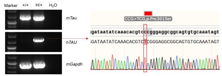
- mRNA expression analysis: Mouse Tau mRNA were detectable in wild-type C57BL/6 mice and heterozygous B-hTAU*P301S mice, Human TAU mRNA was detectable only in heterozygous B-hTAU*P301S mice but not in wild-type mice. Sequence assays showed the heterozygous B-hTAU*P301S mice consists of a C to T point mutation at amino acid 301.
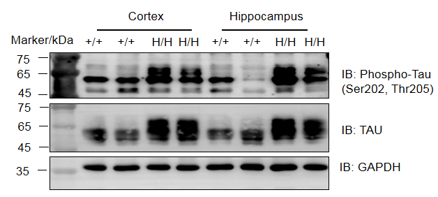
- Protein expression analysis: TAU and p-tau were detected in cortex and hippocampus of both wild-type C57BL/6 mice and homozygous B-hTAU*P301S mice. Additionally, homozygous B-hTAU*P301S mice showed a significant increase in TAU and p-tau levels compared to wild-type C57BL/6 mice.
- Application: This product is used for pharmacodynamics evaluation of Alzheimer's disease (AD).
mRNA expression analysis in B-hTAU*P301S mice

Strain specific analysis of TAU mRNA expression in wild-type C57BL/6 mice and heterozygous B-hTAU*P301S mice by RT-PCR. Brain RNA were isolated from wild-type C57BL/6 mice (+/+) and heterozygous B-hTAU*P301S mice (H/+), and then cDNA libraries were synthesized by reverse transcription, followed by PCR with mouse or human TAU primers. As control, a group without reverse enzyme was also set. Mouse TAU mRNA was detectable both in wild-type C57BL/6 and heterozygous mice. Human TAU mRNA was detectable only in heterozygous B-hTAU*P301S mice but not in wild-type mice.

Western blot analysis of phosphorylation of TAU protein expression in homozygous B-hTAU*P301S mice. Cortex and hippocampus lysates were collected from wild-type C57BL/6J mice (+/+) and homozygous B-hTAU*P301S mice (H/H, 12-week-old mice, n=2), and then analyzed by western blot (Phospho-Tau (Ser202, Thr205) antibody: Invitrogen, MN1020; anti-TAU antibody: CST, #46687). 20 μg total proteins were loaded for western blotting analysis. TAU and p-tau were detected in cortex and hippocampus of both wild-type and homozygous mice.
Histopathological analysis

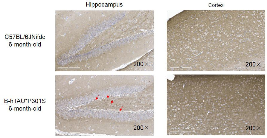
Histopathological analysis of brain in homozygous B-hTAU*P301S mice. The expression of P-TAU in the cortex and hippocampus of 6-month-old C57BL/6J mice (+/+) mice and homozygous B-hTAU*P301S mice was detected by IHC (Phospho-Tau (Thr205): CST, #49561). Compared to wild-type mice, the phosphorylation level of homozygous B-hTAU*P301S mice in the hippocampus was increased, with a small amount of neurofibrillary tangles observed.


 京公网安备: